
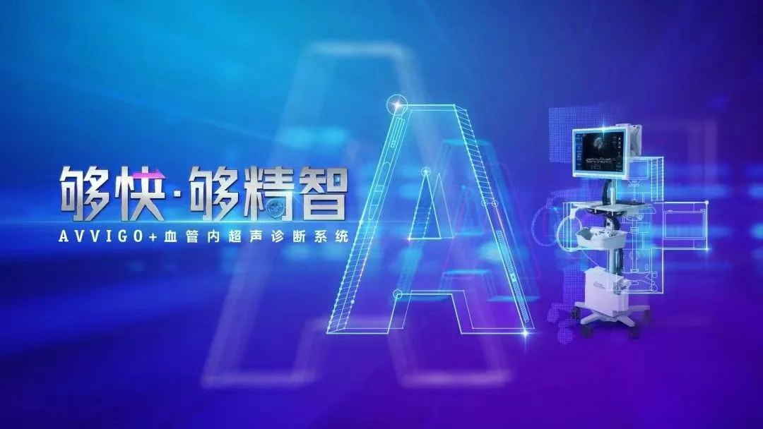
Recently, Professor Mei Weiyi and his team at Guangzhou United Family Hospital successfully completed the center’s first application of the AVVIGO+ Intravascular Ultrasound (IVUS) Diagnostic System. The procedure involved lesion expansion and recanalization for a patient with left coronary artery stenosis and calcification. Leveraging the AVVIGO+ system, the surgical team gained precise insights into the characteristics and details of the diseased vessels, enabling them to select the most appropriate intervention. The operation was a complete success. The AVVIGO+ system empowers precision diagnosis and treatment, optimizes clinical workflows, and significantly improves the outcomes of interventional procedures.
[在此处插入图片 2:AVVIGO+ 系统设备图]
The application of AVVIGO+ IVUS creates a new paradigm for precision coronary intervention. Through real-time image navigation and AI-assisted vascular assessment, it breaks the technical barriers of traditional complex lesion treatments. The system provides fast and efficient vessel analysis results to assist surgeons in decision-making. The new “High-Speed Pullback” feature achieves speeds of up to 8 mm/s, significantly shortening PCI time, reducing ischemia risk in high-risk cases, and greatly enhancing surgical efficiency. AVVIGO+ empowers the diagnosis and treatment of complex lesions with its “fast, precise, and intelligent” capabilities.
Patient Information
- Subject: 56-year-old male foreign national.
- Chief Complaint: History of two previous coronary angiographies indicating atherosclerosis and mild-to-moderate stenosis; long-term medication use. A CTA scan three years ago abroad showed mild stenosis in the Left Anterior Descending (LAD) artery and 70% stenosis in the Diagonal Branch (D1).
- Family History: Both father and younger brother have coronary heart disease with implanted stents.
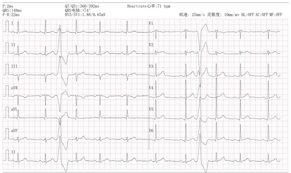
- ECG Findings: Premature Ventricular Contractions (PVCs) and bradycardia.
- Holter Monitor: Sinus rhythm (avg 59 bpm), PVCs (5930/24h), and short runs of Ventricular Tachycardia (1 burst, 3 consecutive ventricular beats).
Pre-operative Angiography
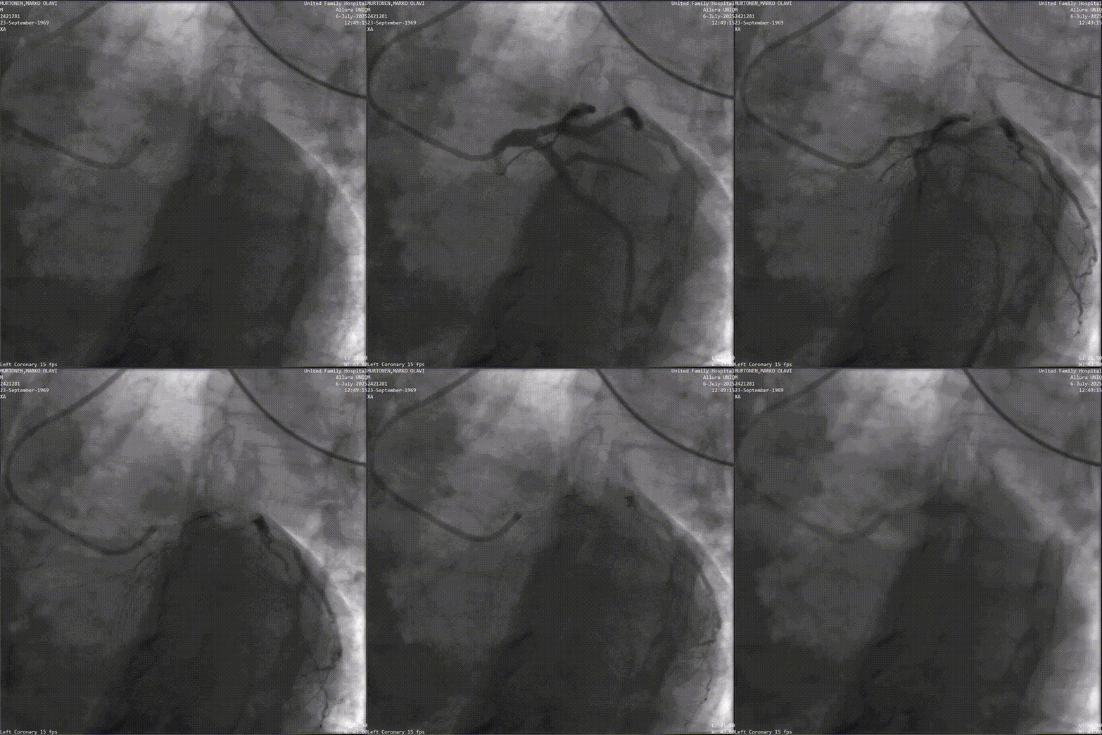
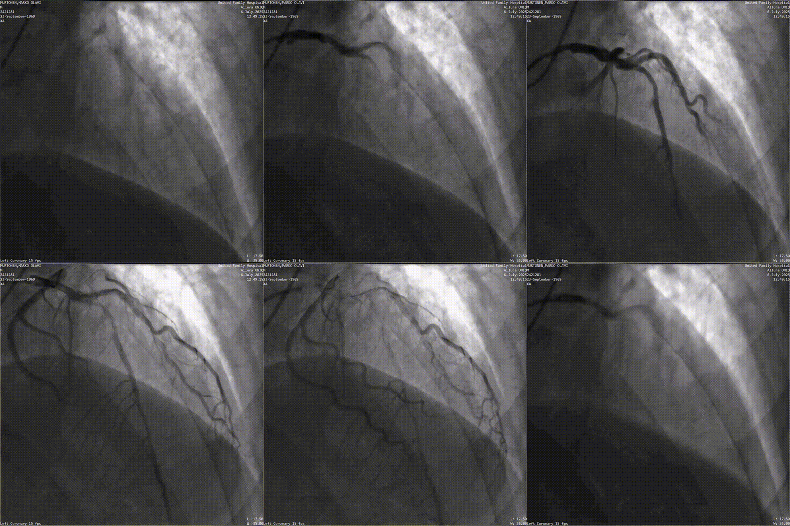
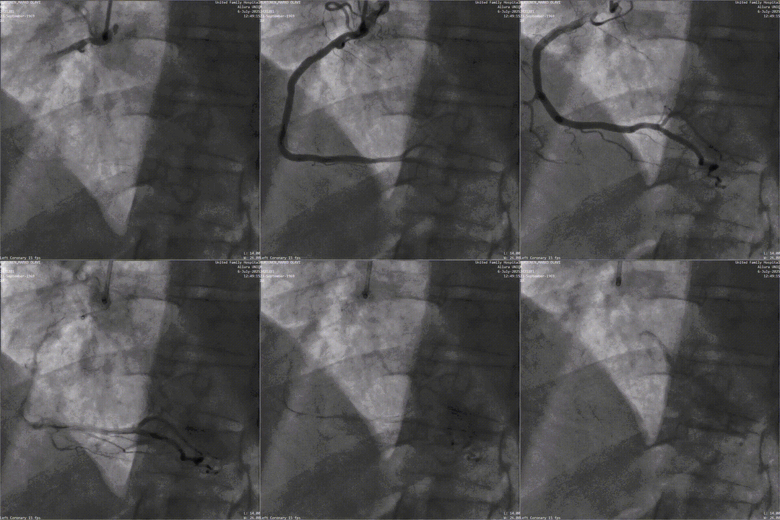
- Right Coronary Artery (RCA): Approximately 50% stenosis in the proximal segment; TIMI Grade 3 forward flow.
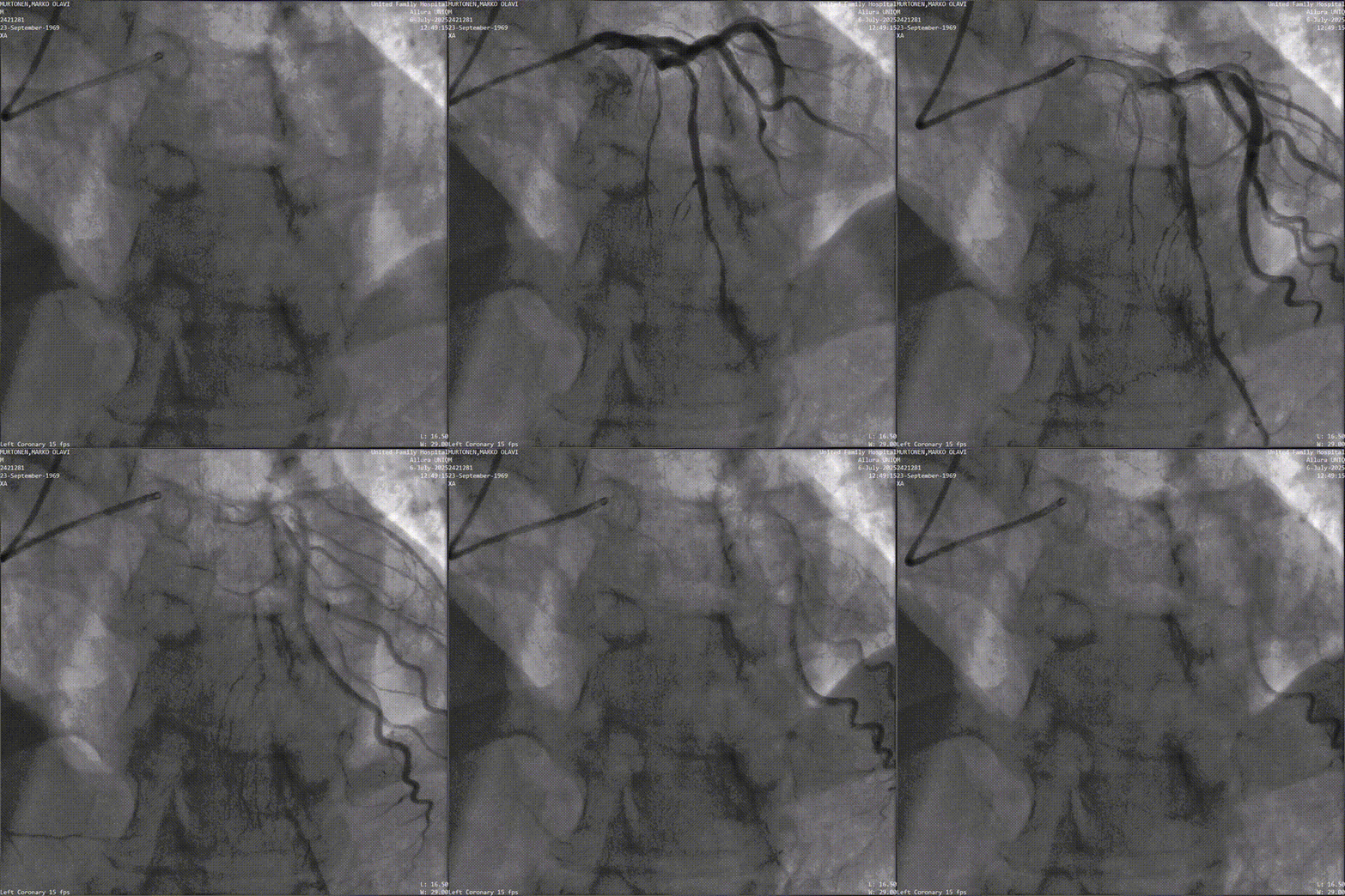
- Left Anterior Descending (LAD): Approximately 60% stenosis with a long lesion; the large Diagonal Branch (D1) has approximately 70% stenosis; TIMI Grade 3 forward flow.
Surgical Procedure
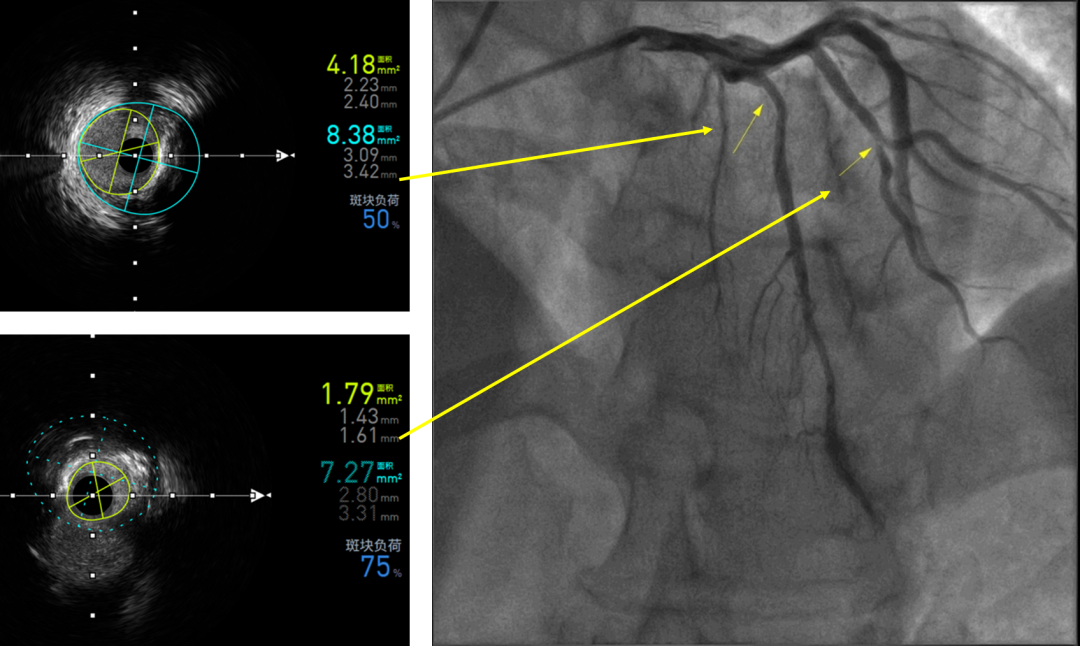
Following the Coronary Angiography (CAG), the surgeon inserted the IVUS catheter and used the AVVIGO+ system to perform endovascular imaging of the LAD-LM and D1-LAD lesions.
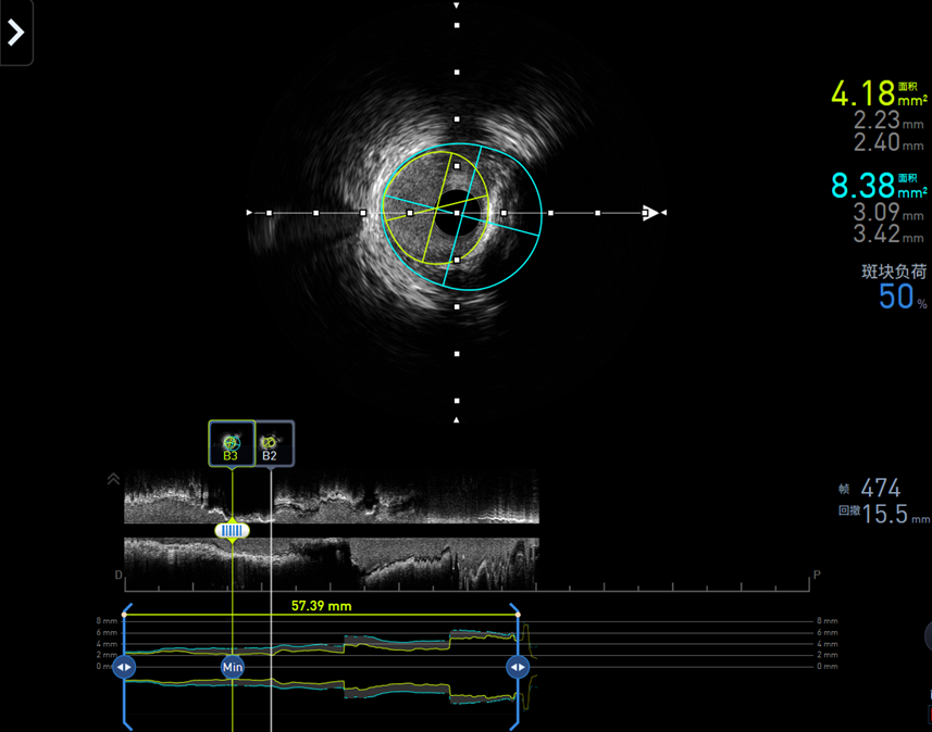
- LAD-LM Analysis: Soft plaques and localized calcified plaques were observed. The Automated Lumen Analysis (ALA) for the proximal LAD showed a Minimum Lumen Area (MLA) of 4.18 mm² and a plaque burden of 50%.
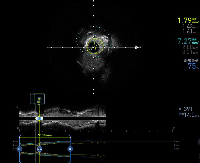
- D1-LAD Analysis: Calcified plaques were seen involving the ostium (opening). ALA showed an MLA of 1.79 mm² at the D1 ostium and proximal segment, with a plaque burden of approximately 75%.
Decision-Making:
Based on the IVUS results and clinical standards, the team determined that the LAD lesion did not require further expansion or stenting; strengthened medication therapy would suffice. However, the D1 lesion met the criteria for intervention. Given the small vessel diameter and the convergence of a large vessel at the stenosis, a Drug-Coated Balloon (DCB) approach was chosen.
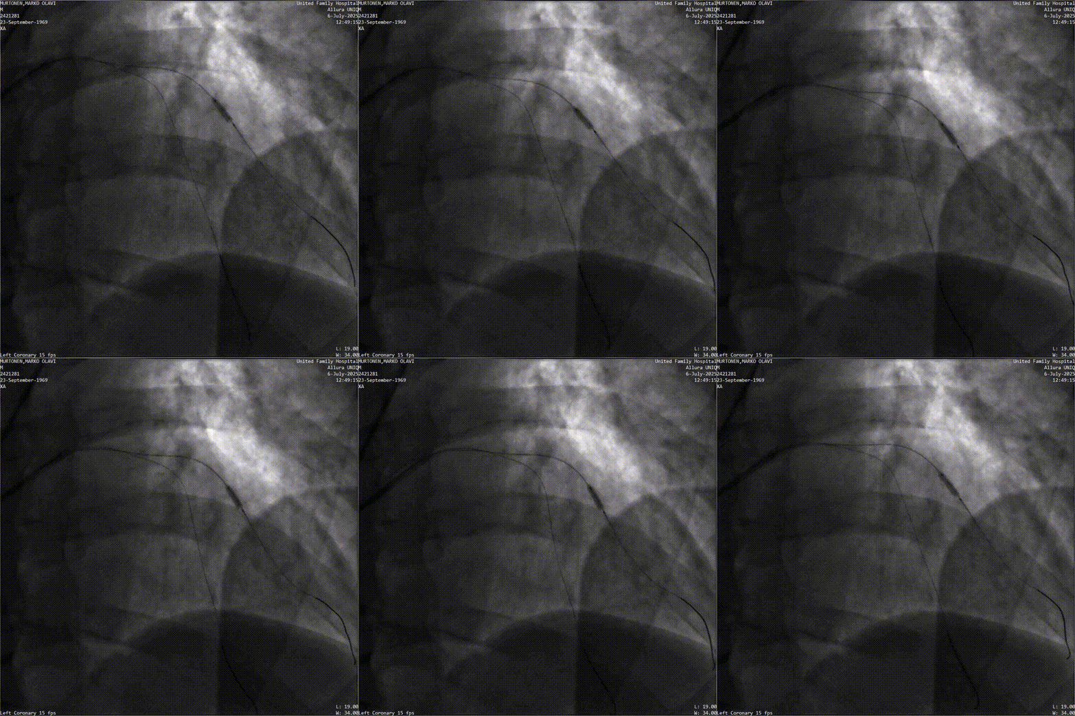
- A 2.25*10mm cutting balloon was used to expand the target lesion twice at 8 atm to gain more lumen area.
- A 2.5*20mm drug-coated balloon was then used to expand the target lesion at 6 atm for 60 seconds.
Results & Follow-up
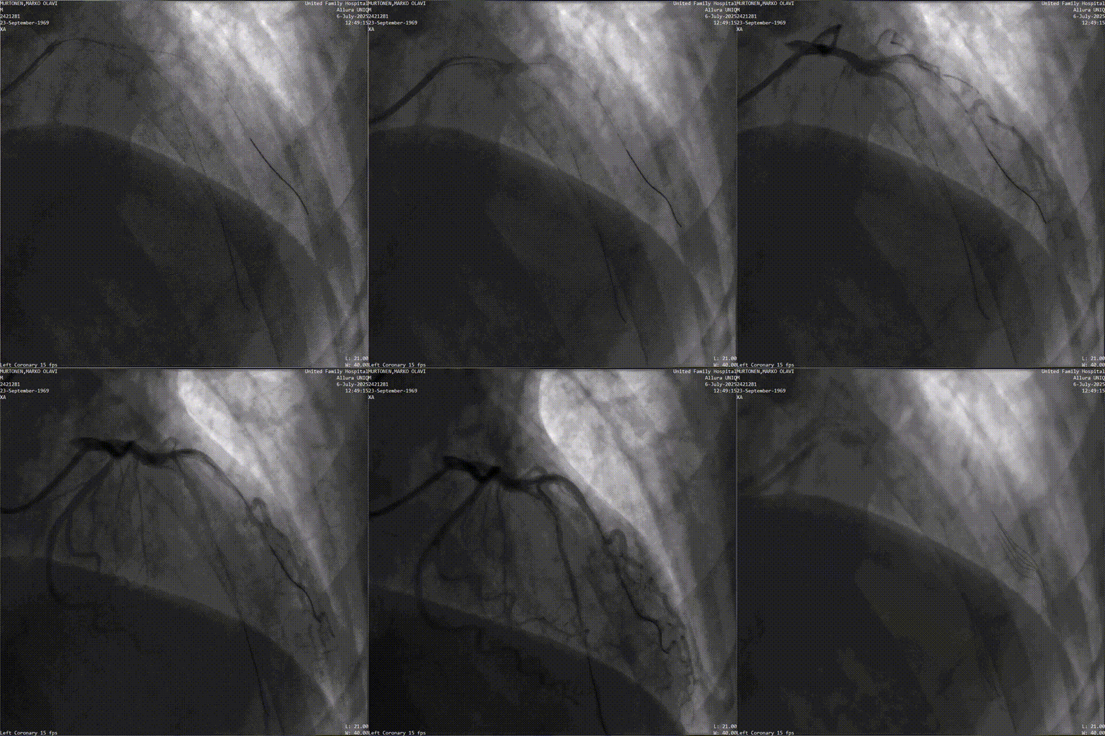
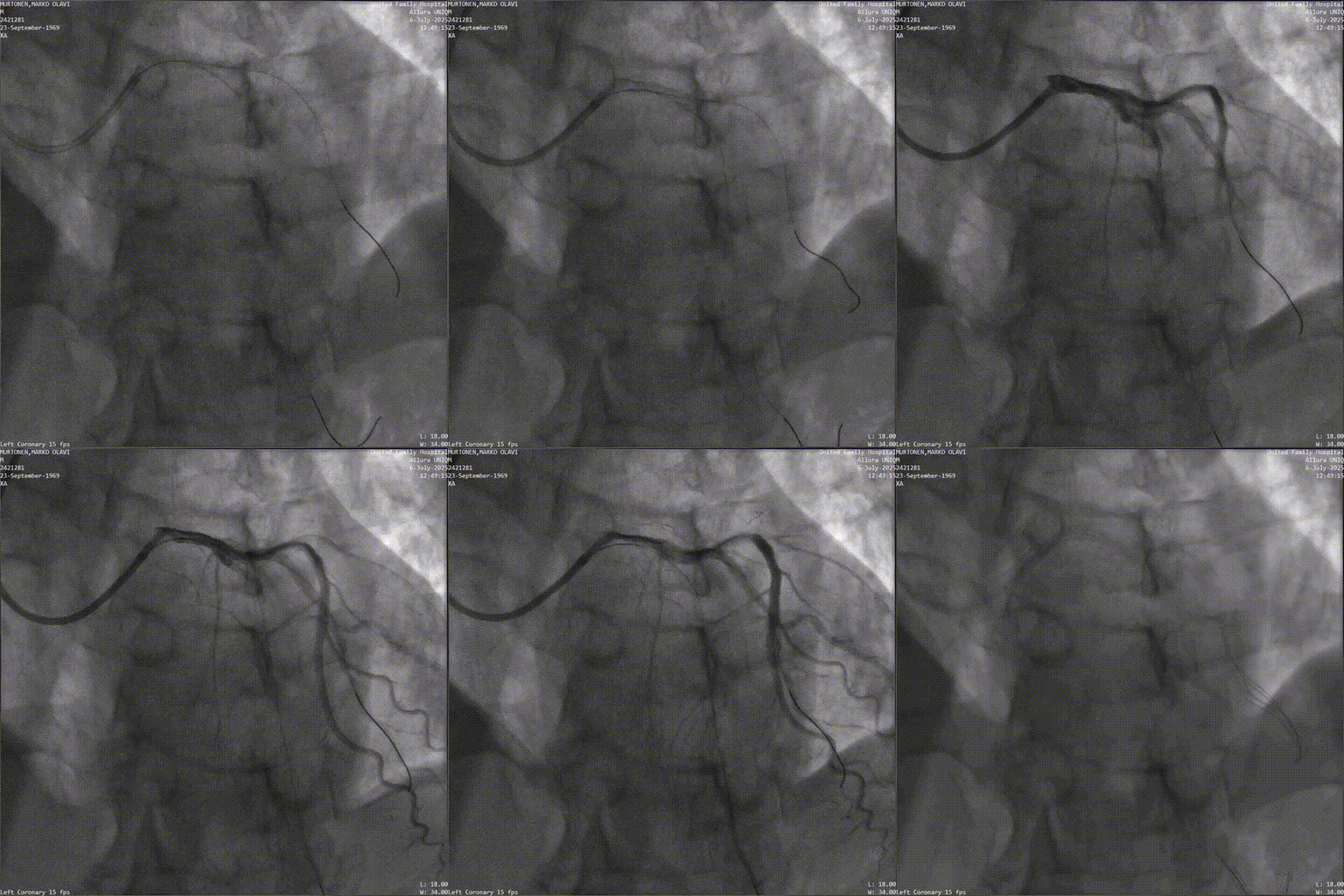
Post-operative angiography confirmed unobstructed distal blood flow, marking a successful surgery.
Post-op Medication: Ticagrelor, Aspirin, Atorvastatin + Ezetimibe, Betaloc-ZOK 11.875mg qd, and Shensong Yangxin Capsules.
Follow-up (One week later):
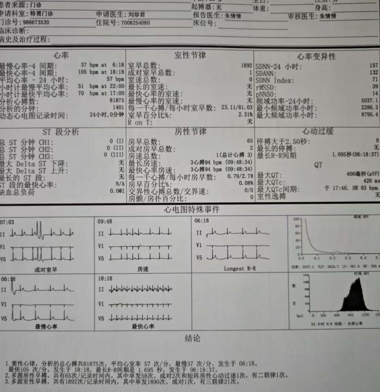
Holter monitor results showed: Sinus rhythm (avg 57 bpm), PVCs reduced to 1892/24h, and no ventricular tachycardia (VT) was observed.
Summary
The AVVIGO+ IVUS system provides a brand-new solution for managing coronary artery lesions. Through high-definition imaging and AI-assisted functions, it provides doctors with real-time, precise endovascular structural information, ensuring accurate placement of balloons and stents, enhancing surgical efficiency, shortening operation time, and reducing complications.
In this case, Professor Mei Weiyi utilized AVVIGO+ to fully master the information of the left coronary lesions and plaque characteristics. Guided by this data, the team successfully implemented a Drug-Coated Balloon (DCB). The reference data provided by AVVIGO+ helped the surgeon achieve “Intervention Without Implantation” (avoiding permanent stents) while ensuring optimal results.
Following the procedure, Professor Mei also experienced the High-Speed Pullback feature. Even at the maximum speed of 8 mm/s, the system maintained exceptional image quality and clarity. Professor Mei expressed high satisfaction with the ALA results and image quality, praising the system’s potential to optimize workflows and safeguard surgical quality.